

Immuno BlootingTechnique antibiotics are used to identify target proteins among unrelated protein species. Via antigen-antibody reactions, protein targets can be identified. Electrophoresis and transfer onto membrane are two techniques used to identify proteins. It is a visualization of specific DNA, RNA, and protein among thousands of contaminating molecules that requires uniformity of number. In this technique, proteins have been separated. Currently in the immunoblotting technique non non-isotopic labels are benign used.
ELISA:
The full form of ELISA is an enzyme-linked immunosorbent assy. It is an immunochemical test that involves enzymes. Antigen or antibodies are also involved in addition to enzymes. It is specifically based on antigen-antibody interaction. ELISA can be used for substances having antigenic properties that can include hormones, bacterial antigens, and antibodies. The antigen is recognized by the specific antibody. This antibody is attached by a second antibody which has an enzyme attached thus it is called enzyme-linked.
Types of ELISA:
There are four types of ELISA methods.
1. Direct ELISA:
In this method of ELISA antigens to be detected are coated directly and nonspecific sites are blocked. Antibody labeled with enzyme is added. Later, to remove unbound antibodies it is washed. Later, the addition of substrate for enzyme and color appears where the amount of antigens are present.
An example of direct ELISA is the detection of urinary beta hCG.

3. Sandwich ELISA:
It is used to detect antibodies and antigens In a well antigen is coated. A sample containing a specific antibody is measured. Wash to remove unbounded antibodies. Against primary antibody, a secondary antibody is added with that substrate for the enzyme is added. A proposition to the amount of antibody present color is sown.
An example of Sandwich ELISA is the detection of anti-HBV antibodies in serum.
Variants of sandwich ELISA:
- Double antibody sandwich ELISA
- Triple antibody sandwich ELISA
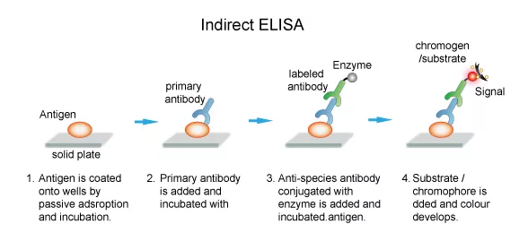
3. Indirect ELISA:
Indirect ELISA is the opposite of direct ELISA. In ELISA antigen is well coated and antibody detects in serum.
4. Competitive ELISA:
In a sample containing antigen, an antibody is incubated in the solution. In an antigen-coated well that mixture of antigen-antibody is added. In the sample more antigen is present, and less amount of antibody is available to bind to the antigen-coated well. Give that solution wash to remove the Ag-Ab complex. Against primary antibody secondary antibody is added. A substrate for the enzyme is added. Present of color is inversely proportional to the amount of antigen present.
Application of ELISA:
- Detection of antigen:
- To detect Infection.
- To detect hormones.
- To detect cytokines.
- Detection of antibody:
- To detect infection
- To detect autoimmunity
Western blotting:
It is used to detect proteins. The basic principle of western blotting is separating proteins into polyacrylamide gel according to the isoelectric point and molecular weight. It is an analytical technique used in molecular biology. Immunoblottings are employed in diagnostic laboratories to identify the desirable protein antigens in the complex mixture. In this technique, proteins are separated by electrophoresis.
Key elements for this technique are:
- Separation
- Efficient transfer
- Specific detection
The procedure of western blotting:
- Load and separate protein samples on SDE-PAGE.
- Electrophoretically transfer franctioned protein onto PVDF membrane.
- Block the membrane with neutral protein.
- Incubate the membrane with primary antibody.
- Incubate the membrane with a secondary antibody specified to the primary antibody.
- Expose the film by introducing chemiluminescence
Blocking agents used in Western blotting:
- Nonfat milk.
- Casein
- Gelatin
- A dilute solution of tween 20
- BSA
Types of protein detection:
- Chromogenic detection
- Chemiluminescence detection
- Fluorescent detection
- Radioactive detection
Limitation of Western Blotting:
- Very time-consuming and delicate procedure.
- Due to secondary antibodies incorrect labeling of protein can happen.
- For this technique well trained technicians are required.
Southern blotting:
E. M. Southern developed a Southern blot technique in 1975. It is used to detect DNA. This technique is used to identify the location of genes and their sequence. In this blotting technique DNA bands from agarose gel to a membrane. The membrane is kept on the gel and it is soaked because of the buffer. It helps to carry DNA from the gel to the membrane where DNA is bound. The same method can be used to transfer RNA and proteins. The key method is hybridization.
Steps involved in southern blotting:
1. Purification and extraction of DNA from the cell:
Isolation of the DNA which is picked from the rest of the cell. Incubate specimen with detergent for cell lysis. Lysis frees protein and DNA from the cell. Organic extraction is used to remove proteins.
2. Restriction of DNA with the enzyme:
Restriction of DNA with the enzyme.

3. Separate DNA by electrophoresis:

A complex mixture is added to the gel to separate electrophoresis according to size.
4. Denature of DNA:
Denaturation of DNA present in gel with the help of alkali. In this method, double strands of DNA are converted into single strands.

5. Transfer those to the nitrocellulose paper:
Transfer of the DNA from the gel to solid support.
6. Wash the unbound probe:
Wash the buffer with NACL and detergent to wash away probes and reduce background.
Disadvantages of southern blotting:
- More expensive than other techniques.
- It is more time-consuming.
Application of Blotting:
- Analysis of IgG fraction purifies from human plasma.
- Diagnosis of HIV.
- A western blotting technique is used to detect Lyme disease.
- A confirmative test for Hepatitis B is done by western blotting.